Scientists just built a 3D atlas for the human body, and it's just like the Google Maps
The 3D atlas is completely free, publicly accessible, and needs absolutely no subscription, institutional login, etc.

Scientists have developed a new tool called the "Human Organ Atlas," allowing users to navigate through real human organs in three dimensions, StudyFinds reported on March 12. Developed by researchers at University College London and the European Synchrotron Radiation Facility in France, the online atlas works just like your Google Maps, but for the human body. But what's so special about it? Well, the 3D atlas is completely free, publicly accessible, and needs absolutely no subscription, institutional login, etc.

As per reports, anyone training in artificial intelligence (AI) systems can access the Human Organ Atlas. The groundbreaking project was published in Science Advances and provided hundreds of detailed organ scans that were previously limited to a few research laboratories.
Now, you must be thinking, why do we even need a 3D atlas if we already have the option of a CT scan to view detailed, cross-sectional, 3D-like images of internal body structures? Well, you're right, but the information provided by a CT scan is far lower in detail than the new atlas.
In fact, while a hospital CT scan captures an entire organ with limited detail, and a microscope provides very fine details in thin, flat slices, the Human Organ Atlas uses hierarchical phase-contrast tomography (HiP-CT), which allows imaging across all scales — from whole organs to near-cellular detail — in 3D and within a single intact organ.

To obtain such specific, detailed scans, scientists use a synchrotron, a machine that makes very precise X-rays, far stronger than what a hospital machine can produce.
At the European Synchrotron Radiation Facility, human organs donated after death and with consent are preserved and scanned. Notably, they first do a full-organ scan at a resolution of about 20 micrometers per voxel. Later, they scan selected regions at much higher resolutions, such as 0.65 micrometers. This process is a bit time-consuming as each scanning session can stretch between two and eight hours, depending on the organ size.
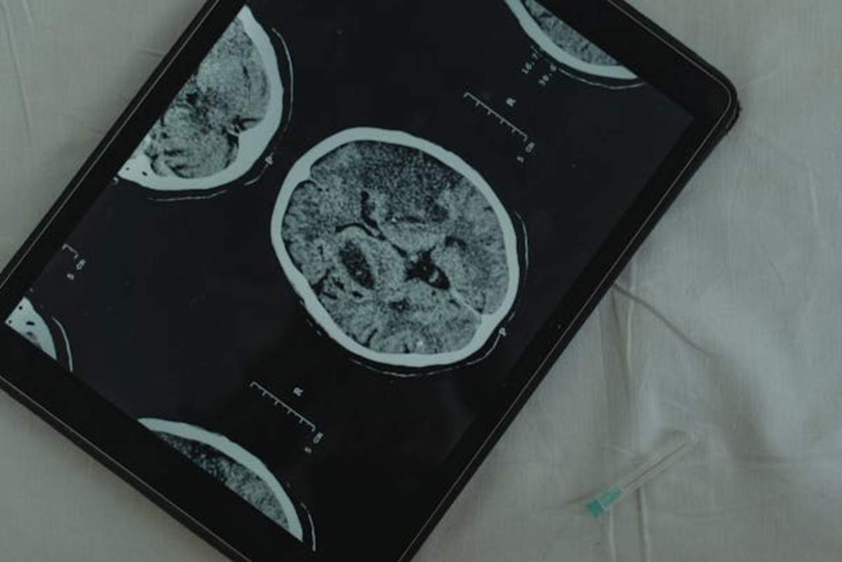
As of March 12, the atlas includes 298 3D image sets obtained from 24 donors (age between 30 and 94), and 10 different organ types — brain, colon, heart, kidney, liver, lung, prostate, spleen, testis, and uterus.
Every year, many people die because of incorrect or late diagnosis. While sometimes the doctors are genuinely at fault, limitations in available tools also play a significant role.
A shocking report published in BMJ Quality & Safety revealed that 371,000 people die every year in the USA following a misdiagnosis, and an estimated 424,000 are left disabled because of it. "Just 15 diseases account for about half of all serious harms, so the problem may be more tractable than previously imagined," the study observed. But this might see a change if the 3D atlas is used more in the medical field.
Besides aging organs, the 3D atlas also has organs with various medical conditions. For example, many donors had passed away during or after the COVID-19 pandemic, and their organs showed evidence of hypertension, cancer, and COVID-19 damage. "Rarer pathologies are represented too, among them a case of Dandy-Walker syndrome, a congenital brain malformation affecting fewer than one in 30,000 people," StudyFinds mentioned in its report.
Now that the 3D atlas is here, we have a different way to study anatomy. Unlike textbooks or cadaver labs, the new tool offers real human organs with diseases in full three-dimensional detail. A student, for instance, can look at a real heart in 3D on a computer. They, in fact, can rotate it to view it from different angles and, if necessary, can also zoom in to look closely at a part, like a valve or the fibers inside a tissue.
More on Scoop Upworthy
Indian teen's invention set to evolve caregiving for Alzheimer's patients in the world
Scientists develop method to implant 3D-printed stem cells as a potential cure for brain injuries
An immigrant couple's quest to cure cancer led to the invention of the Coronavirus vaccine
 Share on Facebook


